Case and discussion contributed by: Omar Al-Nourhji, MD, FRCPC Assistant Professor Department of Pathology and Laboratory Medicine University of Ottawa
James Macpherson, MD General Pathology Resident Department of Laboratory Medicine and Pathology University of Saskatchewan
The following images are representative of a Pap test obtained from a 53-year-old female with complaints of vaginal discharge.
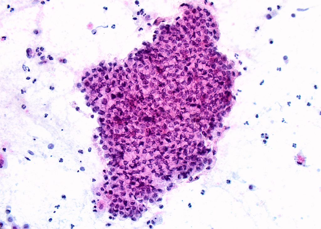
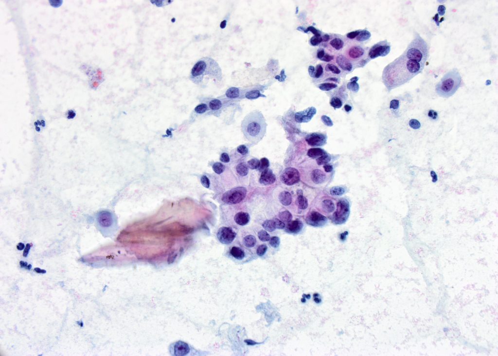
A large sheet of glandular epithelial cells with a mildly disorganized honeycomb pattern. The individual cells demonstrate cytoplasmic mucin and eccentrically located nuclei with mild nuclear atypia.
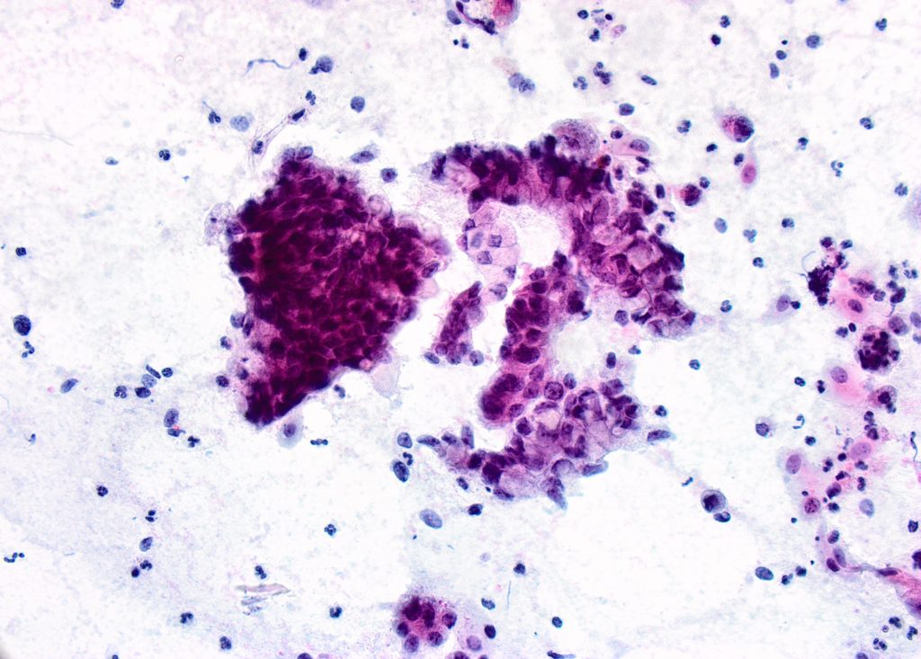
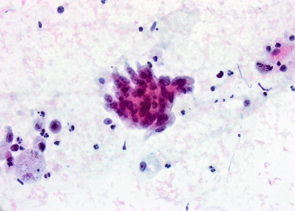
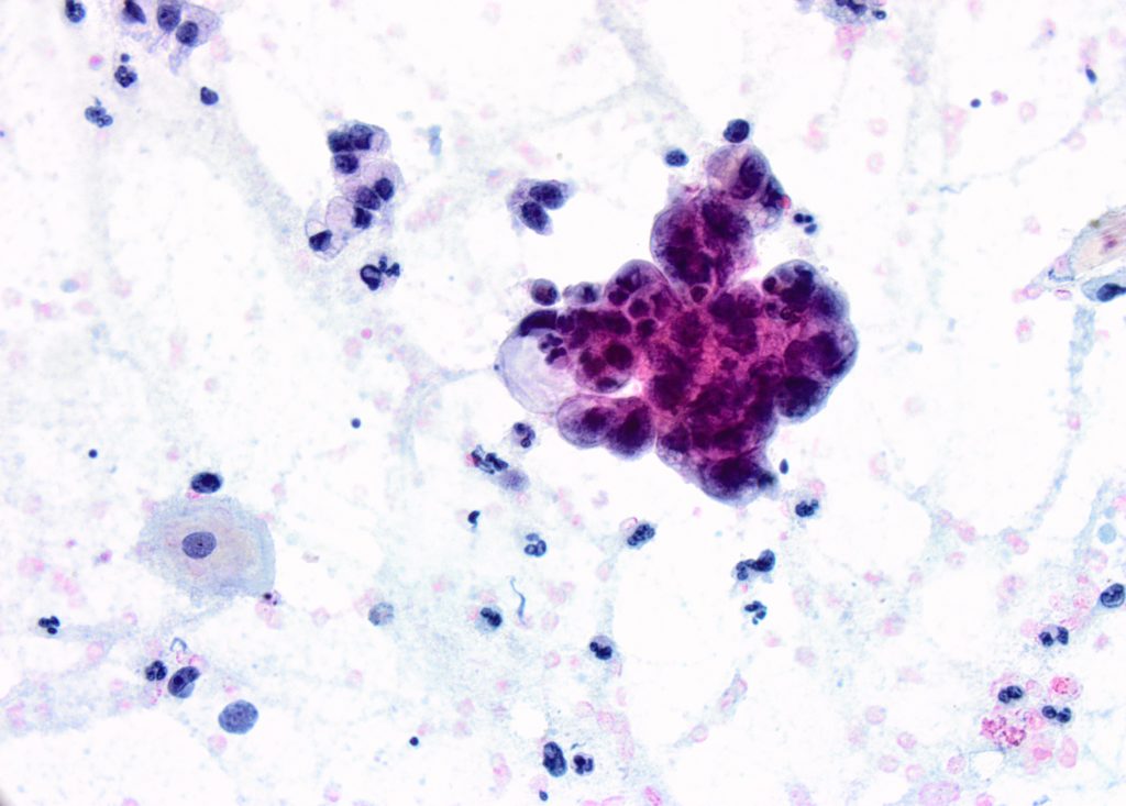
Atypical glandular cells arranged in 3-dimensional group, disorganized sheet/strip and occasional single cells. The individual cells demonstrate cytoplasmic mucin and greater degree of nuclear atypia.
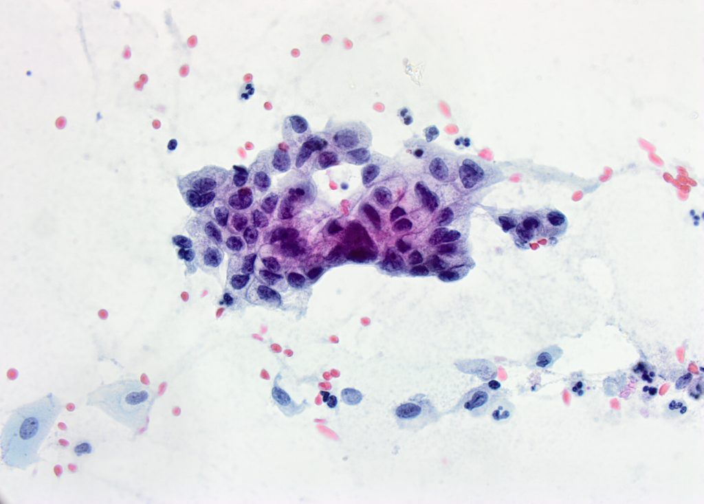
Glandular cells with cytoplasmic mucin, pleomorphic, irregular nuclei and occasional prominent round nucleoli.Cluster of atypical glandular cells with overlapping hyperchromatic nuclei, irregular chromatin distribution and prominent nucleoli.Cluster of atypical glandular cells with clear cellular outlines, cytoplasmic mucin, variation in nuclear size and prominent nucleoli.Occasional groups demonstrated intracytoplasmic neutrophils reminiscent of “bags of polys” seen with endometrial adenocarcinoma.
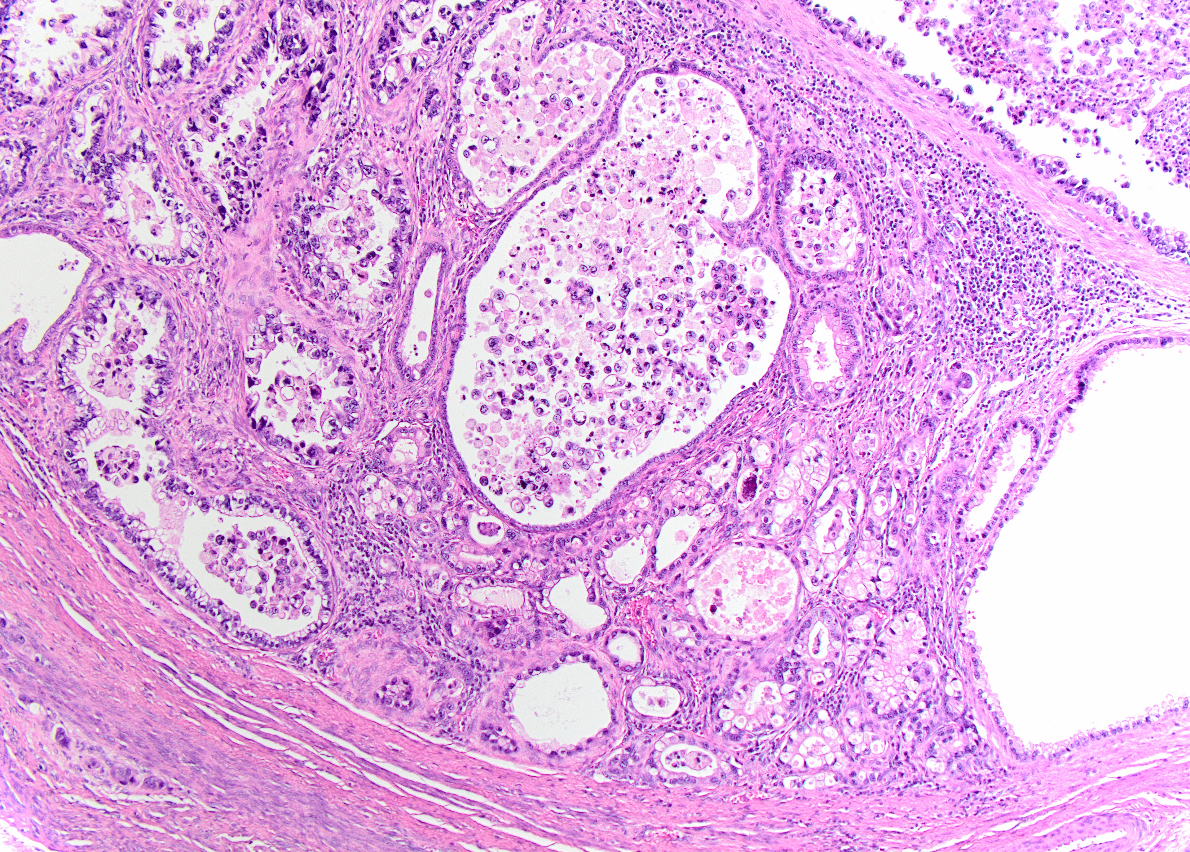
Follow-up surgical resection (H&E 100x) demonstrating infiltrative, irregular glands (endophytic growth pattern) lined by mucinous, gastric type epithelium with clear to pale eosinophilic and voluminous cytoplasm and distinct cell borders.
Follow-up surgical resection (H&E 100x) demonstrating infiltrative, irregular glands (endophytic growth pattern) lined by mucinous, gastric type epithelium with clear to pale eosinophilic and voluminous cytoplasm and distinct cell borders.
Discussion:
Endocervical adenocarcinoma (ECA) is a heterogenous group of malignant neoplasms originating from the glandular epithelium of the uterine cervix. According to the Bethesda System for Reporting Cervical Cytology (TBS), ECA is diagnosed in the presence of abundant abnormal cells, typically with columnar configuration and feathering in a background of necrotic tumor diathesis 1. The cells are usually arranged singly, in three-dimensional clusters or syncytial aggregates and demonstrate enlarged, pleomorphic nuclei with irregular membrane, irregular chromatin distribution, macronucleoli and finely vacuolated cytoplasm. Fulfilment of these criteria is diagnostic of ECA, the most common type being the so-called usual variant2. Further classification of subtypes is usually made on histologic follow-up, although additional cytological features may suggest one of the less common subtypes.
The case presented demonstrates morphologic features meeting TBS criteria for a diagnosis of ECA. It additionally shows cells arranged in a honeycomb pattern with distinct cell borders, abundant foamy mucinous cytoplasm and intracytoplasmic neutrophil entrapment, features suggestive of gastric mucinous endocervical adenocarcinoma (GAS) 3,4, the second most common type of endocervical adenocarcinoma2,4. Additional cytologic features (not identified in our case) are a prominent mucinous background and a “golden-yellow” appearance of the intracytoplasmic mucin which is considered specific for GAS.
Other entities offered in the differential diagnoses are villoglandular and endometrioid variants of endocervical adenocarcinoma. Cytologically, villoglandular adenocarcinoma present as cellular smears with neoplastic glandular cells arranged in branching fragments or bulbous groups reflective of the exophytic long slender papillae seen on histologic sections2. The background is usually relatively clean due to minimal stromal invasion. The cytologic features of endometrioid endocervical adenocarcinoma overlap significantly with those of endometrial endometroid adenocarcinoma with both entities showing clusters of densely packed, rounded and hyperchromatic small cells, resembling the endometrium.
ECA accounts for approximately 20-25% of cervical malignancies, with recently reported increasing incidence. Unlike cervical squamous cell carcinoma, which is HPV driven in almost all cases, up to 10% of ECA in the Western world appears to be HPV unrelated. This is important to keep in mind as screening moves towards cytology and HPV co-testing (in women 30-64 years of age) and even primary HPV testing. ECA has been recently subclassified according to the International Endocervical Adenocarcinoma Criteria and Classification (IECC) into HPV-associated and HPV-unassociated groups 4-5. The HPV-associated subtypes include: ECA usual and villoglandular variants and mucinous adenocarcinoma of intestinal and signet ring cell types. HPV-unassociated types include mucinous endocervical adenocarcinoma of gastric type (GAS), mesonepheric, clear cell, endometrioid and serous carcinomas.
A wide variety of genetic mutations have been reported in GAS with the genes most involved including TP53, CDKN2A, ERBB2/ERBB3, and STK114. STK11 is of importance because of its relation to Peutz-Jeghers syndrome, a rare autosomal dominant inherited syndrome caused by a germline mutation of the STK11 gene and associated with gynecologic malignancies including ovarian sex cord tumor with annular tubules (SCTAT) and cervical minimal deviation adenocarcinoma (MDA).
References:
Wilbur DC, Chhieng DC, Guidos B, Mody DR. (2015) Epithelial Abnormalities: Glandular. In: Bethesda System for Reporting Cervical Cytology. Nayar R, Wilbur DC (eds). 193-240. Springer, Singapore.
Stolnicu S, et al. International Endocervical Adenocarcinoma Criteria and Classification (IECC): A New Pathogenetic Classification for Invasive Adenocarcinomas of the Endocervix. The American Journal of Surgical Pathology. February 2018. Vol.42(2):214-226.
Kawakami F, et al. Cytologic Features of Gastric-Type Adenocarcinoma of the Uterine Cervix. Diagnostic Cytopathology. 2015. Vol. 43(10):791-6.
Hodgson A, Park KJ. Cervical Adenocarcinomas A Heterogeneous Group of Tumors with Variable Etiologies and Clinical Outcomes. Arch Pathol Lab Medicine. January 2019. Vol. 143: 34-46.
Stolnicu S, Barsan I, Hoang L, et al. International Endocervical Adenocarcinoma Criteria and Classification (IECC): a new pathogenetic classification for invasive adenocarcinomas of the endocervix. Am J Surg Pathol.2018;42(2):214–226.
1
2
3
Current
Review
Answered
Incorrect
Question 1 of 3
1. Question
The MOST LIKELY diagnosis is:
Correct
Incorrect
Question 2 of 3
2. Question
Which of the following is a non-HPV-associated endocervical adenocarcinoma?
Correct
Incorrect
Question 3 of 3
3. Question
Which of the following hereditary syndromes-genetic mutations pairs is associated with cervical carcinoma?
 Canadian Society of Cytopathology