A 7-year-old boy was found to have a right frontal bone mass after sustaining a fall at the gym class. Radiologic evaluation revealed a lytic, poorly defined solid and cystic bone lesion involving the right frontal aspect of the calvarium. The following images are representative of a fine needle aspiration of the cystic component of this mass.
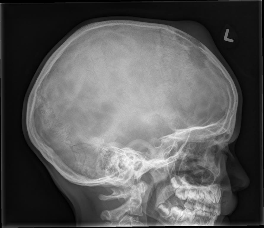
Skull X-Ray: A lytic poorly defined lesion is noted in the right frontal aspect of the calvarium with
associated soft tissue swelling.
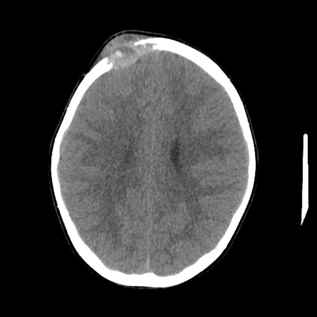
CT scan: Right anterior parietal subcutaneous mass with a fluid level. There is bony destruction of the
underlying calvarium involving the inner and outer tables with irregular moth-eaten margins.
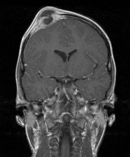
MRI: lytic lesion in the right frontal lobe with a bilobed cystic component and blood/fluid level.
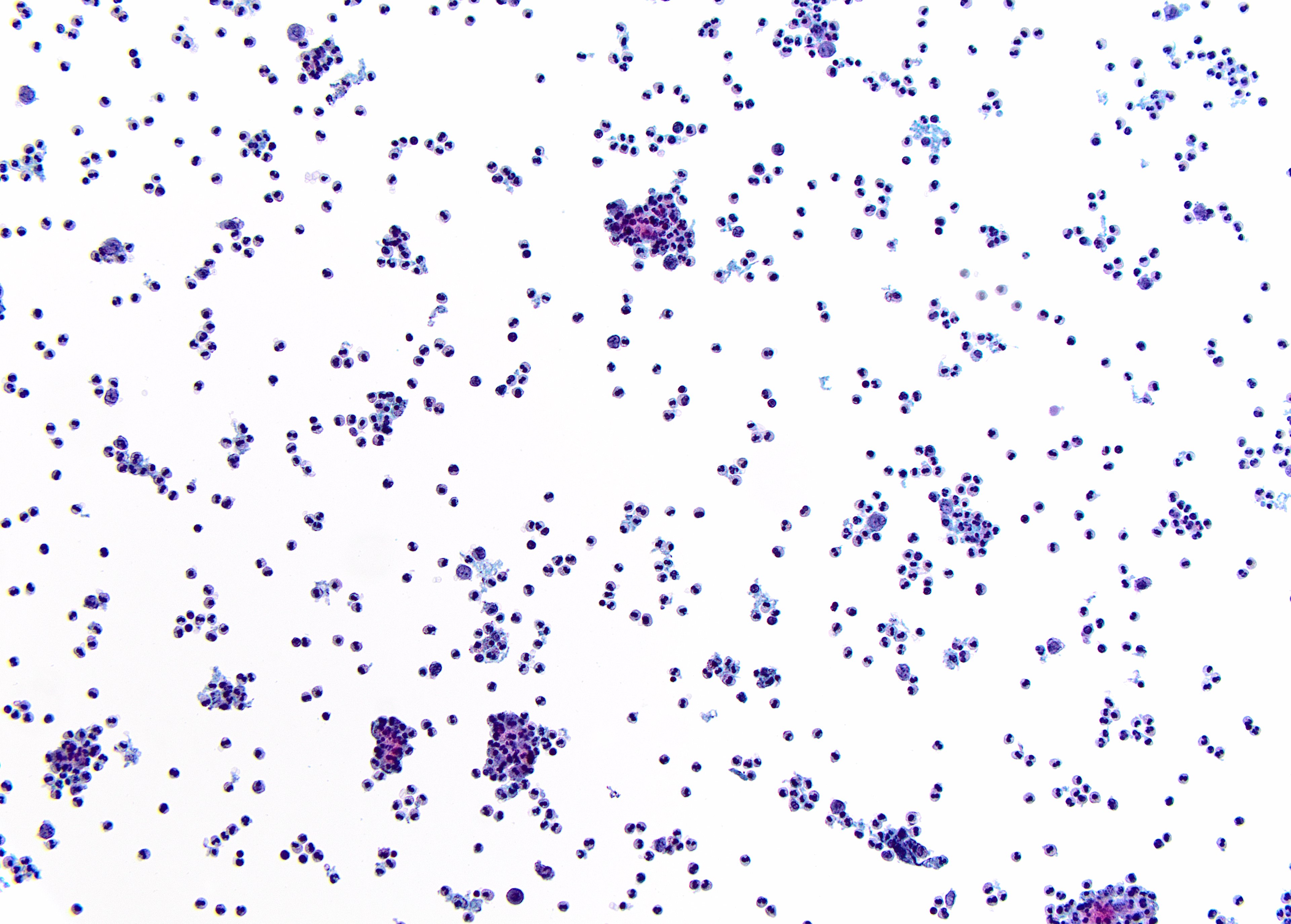
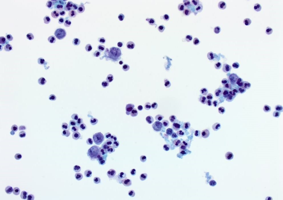
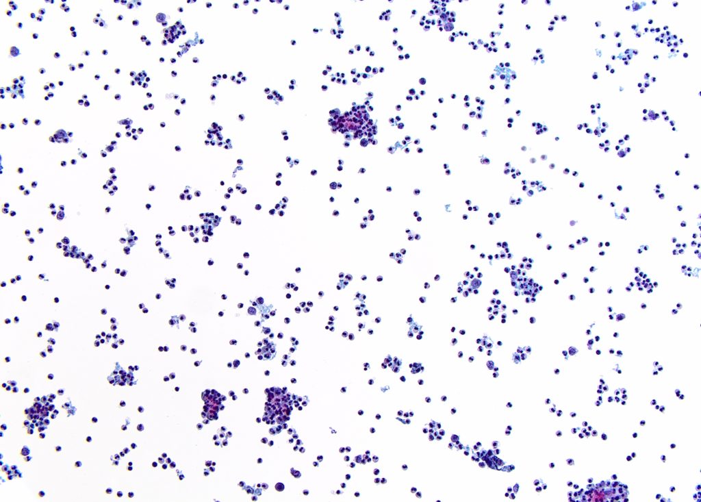
ThinPrep® – Pap stained X20
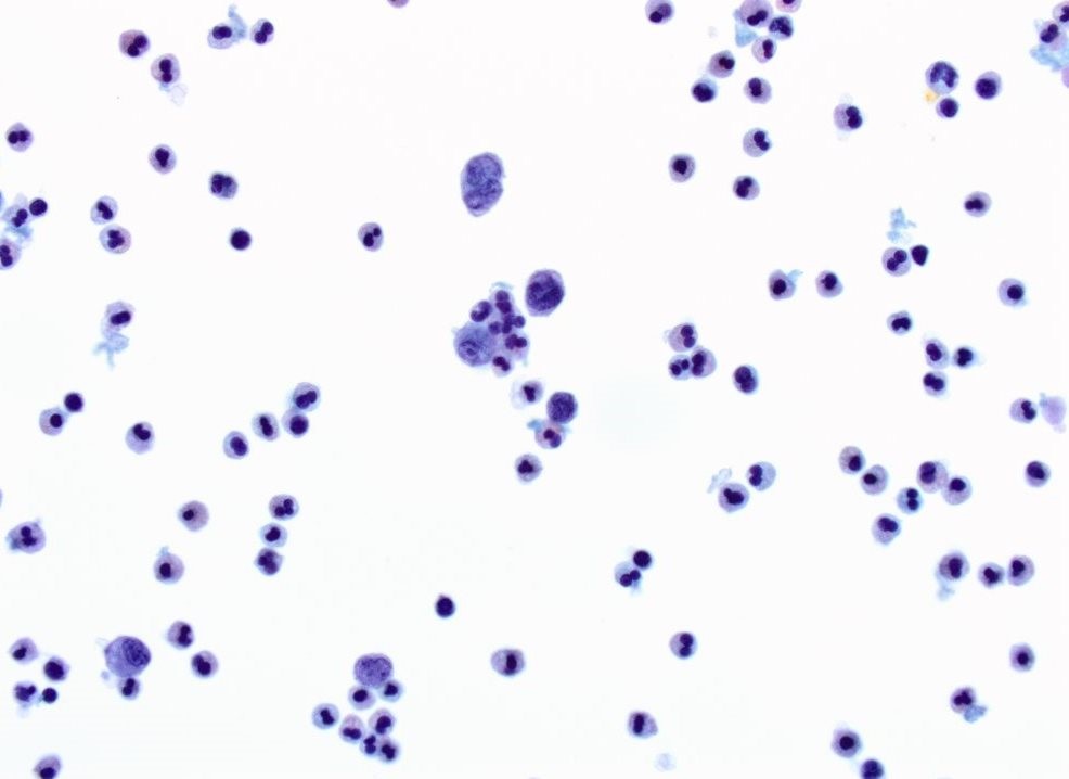
ThinPrep® – Pap stained X60
ThinPrep® – Pap stained X60
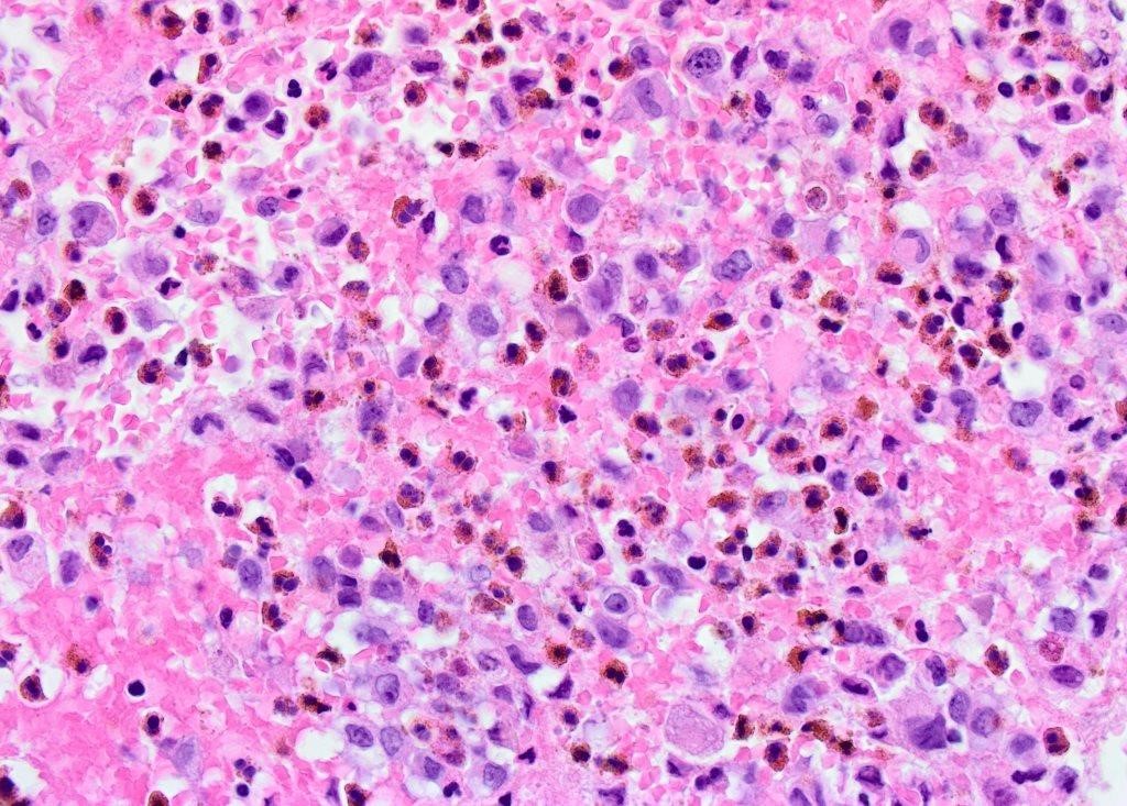
Cell Block – H&E X60
Quiz Summary
0 of 3 questions completed
Questions:
Information
You have already completed the quiz before. Hence you can not start it again.
Quiz is loading…
You must sign in or sign up to start the quiz.
You must first complete the following:
Results
Results
0 of 3 questions answered correctly
Your time:
Time has elapsed
You have reached 0 of 0 point(s), (0)
Earned Point(s): 0 of 0, (0)
0 Essay(s) Pending (Possible Point(s): 0)
Categories
- Not categorized 0%
-
 Canadian Society of Pathology
Case Of The Month
Contributor:
Omar Al-Nourhji, MD, FRCPC
University of SaskatchewanGlenda Wright, MBBCh
General Pathology Resident
Department of Laboratory Medicine and Pathology
University of Saskatchewan
Cellular specimen consistent of scattered epithelioid cells in a background of numerous inflammatory
cells
Higher power view demonstrating neoplastic cells with occasional central nuclear grooves (arrow),
vesicular chromatin and rare small nucleoli. The background inflammatory cells have segmented nuclei
with two to three lobes and no nucleoli.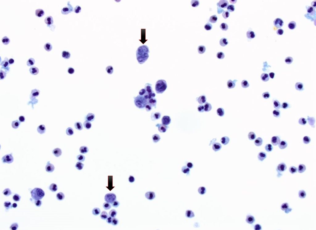
Another higher power view showing neoplastic cells with lobulated nuclei (arrow), vesicular chromatin
and rare small nucleoli.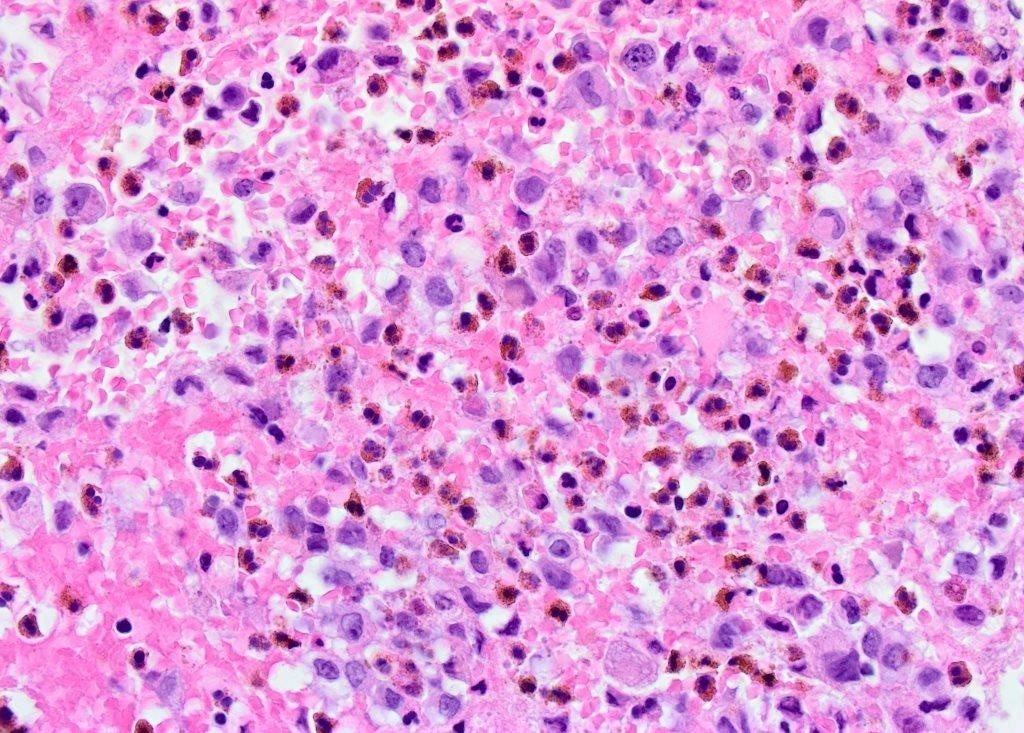
Section of the cell block with large neoplastic cells with ample cytoplasm and complex, folded nuclei
with occasional “coffee bean” grooves and round nucleoli. The background inflammatory cells (better
characterized on H&E) are eosinophils with characteristic segmented nuclei and eosinophilic cytoplasmic granules.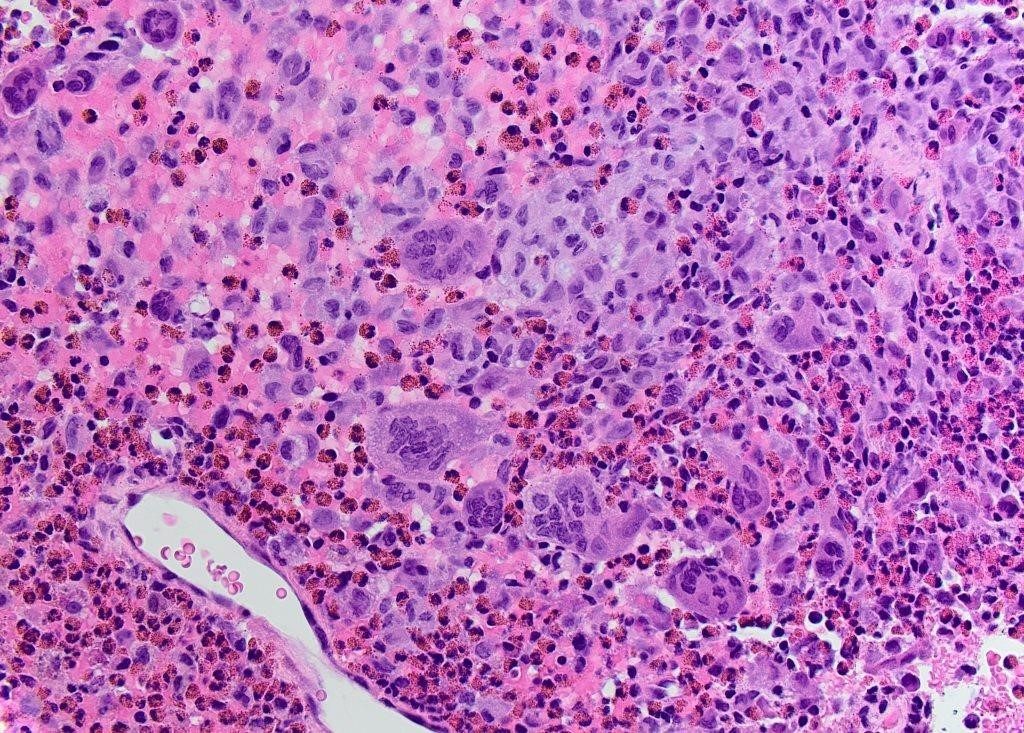
The concurrent biopsy specimen shows similar appearing histiocytes in a background of abundant
eosinophils and multinucleated giant cells.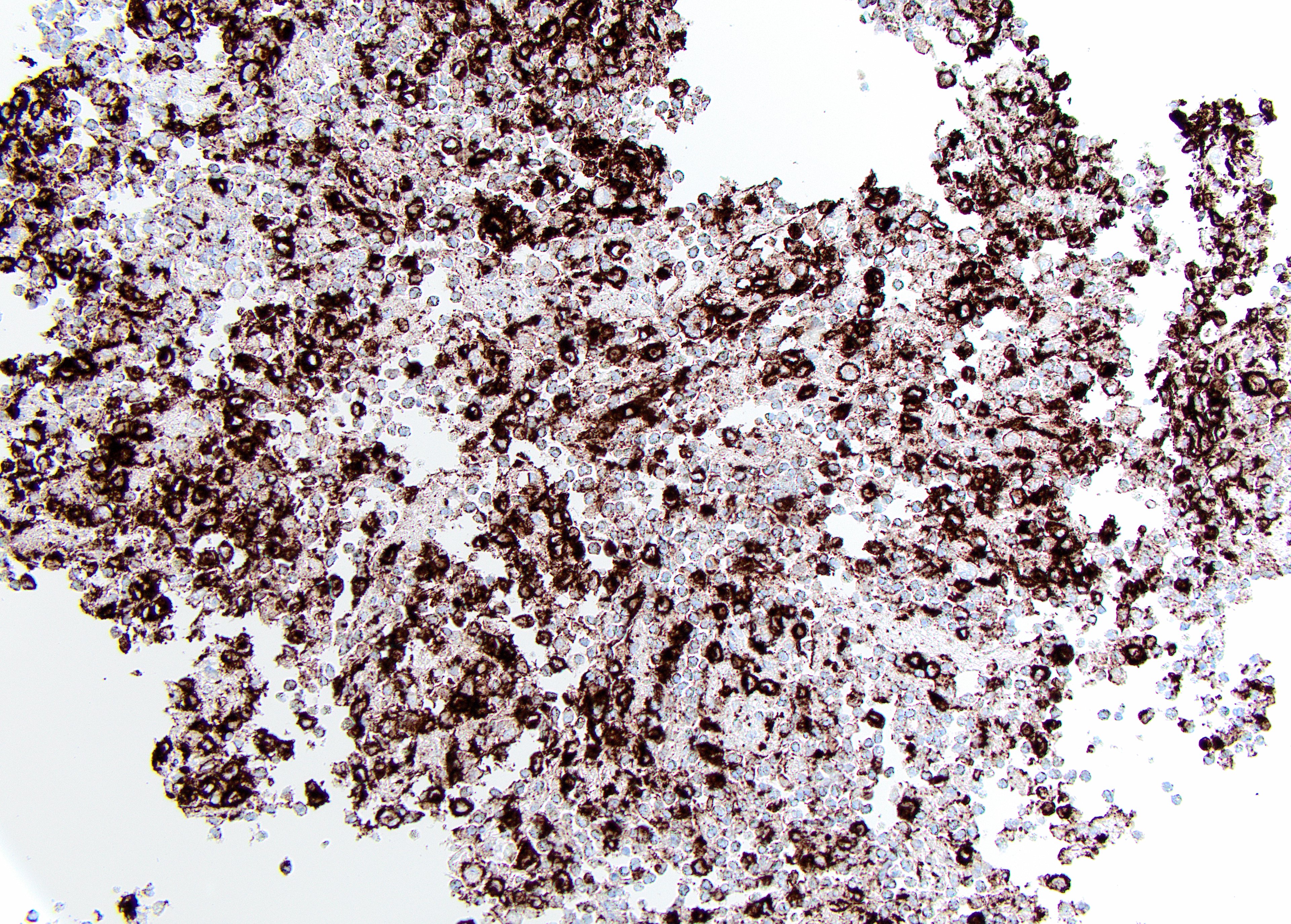
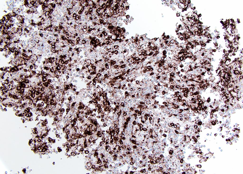
Immunohistochemical staining directed against CD1a is positive within the histiocytes.
Discussion
Based on the patient’s age and radiologic findings, the differential diagnosis in this case includes Ewing
sarcoma (ES), primary lymphoma of bone, Langerhans cell histiocytosis (LCH), aneurysmal bone cyst
(ABC) and osteomyelitis. Cytomorphologic examination of the specimen reveals a cellular aspirate with
polygonal neoplastic cells (Langerhans cells) in a background of prominent inflammatory cells.
Langerhans cells (LCs) are 10–15 µm in diameter and characterized by ample eosinophilic cytoplasm and
lobulated, grooved and indented nuclei with vesicular chromatin and occasional small nucleoli. Thebackground inflammatory cells consist predominantly in this case of eosinophils with multilobed nuclei
(2-3 lobes) and minimal to moderate cytoplasm. They may be confused on initial examination with
neutrophils, due to the absence of the characteristic eosinophilic, granular cytoplasm on monolayer
preparation (ThinPrep®); however, the distinction is very clear upon examination of the cell block. A
concurrent biopsy of the more solid area of the lesion shows LCs surrounded by inflammatory cells,
including multinucleated giant cells. Significant atypia is not identified (excluding Langerhans cell
sarcoma).
Distinction of LCH from other entities listed in the radiologic differential diagnosis is possible based
solely on cytomorphology. Both ES and lymphoma aspirations are cellular with single cell distribution.
The neoplastic cells in both conditions demonstrate a “small blue cell” appearance with round to oval
nuclei, fine chromatin, inconspicuous nucleoli, and scant cytoplasm. A vacuolated tigroid background
maybe appreciated in cases of ES while lymphoglandular bodies maybe noted in lymphoma. Aspirates of
acute osteomyelitis usually consist of abundant neutrophils, macrophages, and necrotic bone. ABC
aspirates are generally paucicellular and blood rich. They may show scattered giant cells, spindle cyst
lining cells arranged singly and in small groups and hemosiderin laden histiocytes.
The characteristic immunophenotype of LCH includes expression of CD1a, S100 protein, and langerin
(CD207) (1) . Expression of CD68 is variable. Ultrastructural examination (electron microscopy) shows the
characteristic Birbeck granules (elongated, zipperlike cytoplasmic structures measuring 200 to 400 nm X
33 nm).
LCH represents a spectrum of idiopathic, neoplastic proliferations of histiocytes (Langerhans cells). It
usually occurs during the 1st three decades of life. It can involve any organ and present as an isolated
focal lesion (monostotic disease), multifocal lesions (polyostotic disease) or as a multi-organ systemic
disease. Localized involvement of the skeletal system is the most common form of LCH and is classically
seen in young children (5-15 years of age). The skull is the most common location with other locations
including the pelvis, spine, mandible and ribs. BRAF V600E mutations have been recently reported in 25-
64% of cases of LCH, resulting in constitutive activation of the mitogen-activated protein kinase (MAPK)
pathway. Mutations in MAP2K1, which encodes the kinase MEK1 protein in the MAPK pathway have
been reported in LCH cases with absent BRAF mutations (4) . Despite these recent discoveries, few
studies have been completed to guide the development of targeted therapy (5) .
The other genetic mutations listed in question 3 are associated with entities included in the differential
diagnosis with EWSR1-FLI1 gene fusion identified in Ewing sarcoma; EWSR1-WT1 gene fusion associated
with desmoplastic small round cell tumor, rearrangement of USP6 locus reported In ABC and IgH gene
rearrangement being a feature of B-cell lymphomas.
References
- Harmon CM and Brown N. Langerhans Cell Histiocytosis: A Clinicopathologic Review and
Molecular Pathogenetic Update. Arch Pathol Lab Med. 2015 Oct;139(10):1211-4 - Haroche J, Charlotte F, Arnaud L, et al. High prevalence of BRAF V600E mutations in Erdheim-
Chester disease but not in other non-Langerhans cell histiocytoses. Blood.
2012;120(13):2700–2703 - Go H, Jeon YK, Huh J, et al. Frequent detection of BRAF(V600E) mutations in histiocytic and
dendritic cell neoplasms. Histopathology. 2014;65(2):261–272 - Brown NA, Furtado LV, Betz BL, et al. High prevalence of somatic MAP2K1 mutations in BRAF
V600E-negative Langerhans cell histiocytosis. Blood. 2014; 124(10):1655–1658 - Allen C., Merad M and McClain K. Langerhans-Cell Histiocytosis. N Engl J Med. 2018 Aug
30;379(9):856-868
* Case and discussion contributed by:
Omar Al-Nourhji, MD, FRCPC
Assistant Professor
Department of Laboratory Medicine and Pathology
University of Saskatchewan - Harmon CM and Brown N. Langerhans Cell Histiocytosis: A Clinicopathologic Review and
- 1
- 2
- 3
- Current
- Review
- Answered
- Incorrect
-
Question 1 of 3
1. Question
The MOST LIEKLY diagnosis is:
CorrectIncorrect -
Question 2 of 3
2. Question
Which immunohistochemical (IHC) stain is expected to be positive in this condition?
CorrectIncorrect -
Question 3 of 3
3. Question
Which of the following genetic mutation is associated with this condition?
CorrectIncorrect